>What does the nervous system due
>Diffusion & Action potentials in Neurons- rapid transmission of messages
>Reflex arc (simple somatic function) and autonomic function
>What can we sense
Function of nervous system
The nervous system has three major functions and consists of two major parts the central nervous system (CNS) and the peripheral nervous system (PNS). The brain and spinal cord and considered the CNS and the nerves throughout the body are the PNS. These two systems work together to perform the following; sensory input, integration, and motor output. When internal or external stimuli is detected by sensory receptors and nerve impulse is generated and travels through the PNS to the CNS. The CNS then takes the information it is receiving and the generates motor out put. Which are more nerve impulses but coming from the CNS to PNS and then specific muscles or organs. For example when you eat food the sensory receptors in the stomach send an impulse through PNS to your CNS saying that there is food in here. Then your CNS sends out an impulse to your stomach, pancreases, liver, gallbladder all the organs associated with digestion to get to work.

Diffusion & Action potentials in Neurons- rapid transmission of messages
Nerve impulses are transmitted throughout the body by cells called neurons. Neurons consist of a cell body, dendrites, and an axon. The nucleus and organelles are located in the cell body. The dendrites receive messages from sensory receptors and other neurons. While the axon conducts nerve impulses. As a nerve impulse occurs a rapid change in polarity across the axonal membrane called action potential takes place. It takes a certain level called threshold for an action potential to occur. An action potential strength stays constant but an intense stimulus can cause the start of an axon potential. Meaning that nerve impulses (action potential) is an either takes place or doesn’t and the intensity of the message is determined by how many nerve impulses take place in a given time frame. Axon length varies for each type of neuron and most are covered in myelin sheath a protective lipid substance. The myelin sheath wraps forms where neuroglia cells called Schwann cells wrap themselves around the axon in many layers. Gaps occur making nodes of Ranvier because neuroglia cells only cover part of the axon. There are also unmyelinated axons which are usually short axons while the long axons have the myelin sheath. Not only does the myelin sheath protect it also acts as a passageway for new fiber growth in nerve regeneration. At the end of an axon many branches with axon terminals on the end extend outward towards another neurons dendrites or cell body. The area between the axon terminals (sending neuron) and the receiving neuron is called a synaptic cleft. Neurotransmitters from the axon of the sending neuron are released crossing the synaptic cleft and bind with the receiving neuron.

Nerve impulse travel at 1.0 m/sec in unmyelinated axons and 100m/sec in myelinated axons and this type of conduction is referred to as salutatory conduction. This is because in myelinated axons a nerve impulse jumps from node to node while unmyelinated axons the action potential (nerve impulse) at one location stimulates an adjacent part of the axon’s membrane to produce an action potential. When people are diagnosed when multiple sclerosis and leukodystrophies, demyelization ,the loss of myelin, has occurred which effects the function of the nervous system.
Reflex arc (somatic function) and Autonomic function
The somatic system has many nerves that serve the skin, skeletal muscles and tendons. These nerves take messages to the CNS from external sensory receptors and then motor commands from the CNS to skeletal muscles. Now while actions are voluntary like walking or eating some are also automatic referred to as a reflex. A reflex occurs when an external sensory receptor is triggered like touching a hot surface. The sensory receptors in your skin send a message to your CNS where a response is then sent back the muscles in your hand and arm to pull away and to feel pain. This happens very fast and without thinking about it resulting in the quick movement known as a reflex. The autonomic system on the other hand is involuntary. After you eat you no longer think about the food or what happens to it but you autonomic system does. It regulates all your organs telling them when to function and when not too. This system is divided into two part the sympathetic division and parasympathetic both consisting of fibers . The sympathetic division is located in the middle portion of the spinal cord and branches out into ganglia, collection of cell bodies outside of CNS) almost immediately. The preganglionic fiber is short while the postganglionic fiber is long and is the fiber that makes contact with an organ. This part of the autonomic system is very important during emergency situations. The sympathetic division accelerates the heartbeat, dilates air passages and inhibits the digestive tract. An example being when walking outside in the dark if you hear something frightening and don’t know what it is your heart starts beating really fast and you feel the need start to run. The sympathetic division has many other function some being; stops tears, dilates pupils, and stimulates the release of glucose from liver. The parasympathetic division includes a portion of the bottom of the spinal cord and a few cranial nerves. Unlike the sympathetic division the preganglionic fiber is long and the postganglionic fiber is short. This is because the ganglia is either near or inside the organ. The parasympathetic division is known as the housekeeper of the rest and digest system. It promotes the responses we associated with rest like slowing the heartbeat and stimulates the organs associated with digestion of food. 
What can we sense?
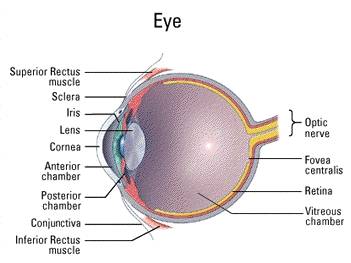
First off the reason why we can sense anything is because we have sensory receptors which are dendrites that are specialized to detect certain stimuli from both internal and external. Exteroceptors sense stimuli from outside the body while interceptors sense stimuli from inside the body. The four types of sensory receptors are chemoreceptors, photoreceptors, mechanoreceptors and thermoreceptors. Chemoreceptors recognize stimuli from taste, smell, and they monitor blood pH. Pain receptors are a type of chemoreceptors that consist of special dendrites that respond to chemicals produced by damaged tissue. Pain receptors also alert us to danger like when touching something sharp or hot. Photoreceptors are stimulated by light energy. Our eyes have photoreceptors both rod cells and cone cells giving us the ability to see black, white and different colors. Mechanoreceptors allow use to hear by meaning of sound waves, sense changes in gravity and motion allowing us to keep our balance, and the sense of touch from pressure receptors. These pressoreceptors detect changes in blood pressure and how much the lungs have inflated. There are also proprioceptors that make us aware of our limbs. Lastly we have thermo receptors that are stimulated my temperature and are located in the skin and hypothalamus. All of these receptors allow us to sense by means of hearing, seeing, taste, touch, and smell. Our nose gives use the ability to smell both good and bad aromas. While the taste buds that cover our tongue allow us to taste the food we are eating. Our ears sense sound waves alerting us and our eyes sense light allowing us to see the world and our skin lets us feel everything we come into contact with. The many different senses and sensory receptors send these impulses to our brain for interpretation so that we may experience what is taking place whether good or bad.


Work Cited:
Madder, Sylvia S. “Human Biology” 10th ed. New York: McGraw-Hill, 2007
http://www.infovisual.info/03/img_en/038%20Nervous%20system.jpg
(Nervous system)
http://mustang.millersville.edu/~bduncan/465/anatomy/brain-anatomy/cellular-level/neuron.gif
(Neuron)
http://www.wooster.edu/psychology/intro/autonomic.gif
(autonomic system)
http://fig.cox.miami.edu/~cmallery/150/neuro/c7.49.3.skin.jpg
(touch)
http://www.rennard.org/alife/img/biom/eye_anatomy.jpg
(eye)
http://www.lincoln.dubuque.k12.ia.us/images/Fifth%20Grade/2004_2005/human_body/tongue.jpg
(tongue)
http://www.hf.faa.gov/Webtraining/NonVisDisplays/Non_vis%20images/earcrop.jpg (Ear)

1 comment:
I likes the clarity of the text and the images were great. Jim
Post a Comment